


Anterior cruciate ligament injury
|
|
|
|---|---|
 Diagram of the right knee
|
Top of page
The combination of "pop" during a twisting movement or rapid deceleration, together with inability to continue participation, and followed by early swelling, is said to indicate a 90% probability of rupture of the anterior cruciate ligament.
Causes

ACL injuries occur when an individual stops suddenly or plants his/her foot hard into the ground (cutting). ACL failure has also been linked to heavy or stiff-legged landing; the knee rotating while landing, especially when the knee is in an unnatural position.
Women in sports such as association football, basketball, and tennis are significantly more prone to ACL injuries than men. The discrepancy has been attributed to gender differences in anatomy, general muscular strength, reaction time of muscle contraction and coordination, and training techniques. Women also have a relatively wider pelvis, requiring the femur to angle toward the knees. This angle towards the knee is referred to as the Q angle. The average Q angle for men is 14 degrees and the average for women is 17 degrees. Steps can be taken to reduce this Q angle, such as using orthotics. The role of genetics is currently speculative.
Significantly many ACL injuries occur in athletes landing flat on their heels. The latter directs the forces directly up the tibia into the knee, while the straight-knee position places the anterior femoral condyle on the back-slanted portion of the tibia. The resultant forward slide of the tibia relative to the femur is restrained primarily by the now-vulnerable ACL.
Ligament dominance
The increased risk of anterior cruciate ligament injury among female athletes is best predicted by the motion and loading of the knee during performance situations. The ligament dominance theory suggests that females typically perform athletic movements with greater knee valgus angles. A greater amount of stress is placed on the ACL in these situations because there is high activation of the quadriceps muscles despite limited knee flexion, limited hip flexion, greater hip adduction, and a large knee adductor moment. Additionally, females typically land with their tibia rotated internally or externally. As a result of increased knee valgus stress, ground reaction forces are greater and laterally directed.
Quadriceps dominance
Ligament dominance is observed when there is excessive movement in the frontal plane to accommodate limited movement in the sagittal plane. This is caused by weakness in the hamstring muscles or reliance on the strength of the quadriceps muscles. This quadriceps dominance theory identifies when the hamstring muscles are notably weaker than the quadriceps muscles. As a result, knee stability in performance situations depends on the quadriceps due to a discrepancy in the pattern in recruiting quadriceps and hamstring muscles.
Trunk and leg dominance
Other theories used to explain the increased risk of ACL injury among female athletes include the trunk dominance and leg dominance theories. Trunk dominance suggests that males typically exhibit greater control of the trunk in performance situations as evidenced by greater activation of the internal oblique muscle. Leg dominance suggests that females exhibit greater kinematic leg asymmetry in knee valgus angles, hip abduction, and ankle abduction in performance situations.
 |
 |
| Right knee, front, showing interior ligaments | Left knee, behind, showing interior ligaments |
Top of page
Diagnosis
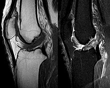
The pivot-shift test, anterior drawer test and Lachman test are used during the clinical examination of suspected ACL injury. The Lachman test is recognized by most authorities as the most reliable and sensitive test, and usually superior to the anterior drawer test. The ACL can also be visualized using a magnetic resonance imaging scan (MRI scan).
An ACL tear can present with a popping sound heard after impact, swelling after a couple of hours, severe pain when bending the knee, and buckling or locking of the knee during movement.
Though clinical examination in experienced hands can be accurate, the diagnosis is usually confirmed by MRI, which has greatly lessened the need for diagnostic arthroscopy and which has a higher accuracy than clinical examination. It may also permit visualization of other structures which may have been coincidentally involved, such as a meniscus, or collateral ligament, or posterolateral corner of the knee joint.
Top of page
Treatment
The term for non-surgical treatment for ACL rupture is "conservative management", and it often includes physical therapy and using a knee brace. Instability associated with ACL deficiency increases the risk of other knee injuries such as a torn meniscus, so sports with cutting and twisting motions are problematic and surgery is often recommended in those circumstances.
Patients who have suffered an ACL injury should be evaluated for other injuries that often occur in combination with an ACL tear and include cartilage/meniscus injuries, bone bruises, PCL tears, posterolateral injuries and collateral ligament injuries.
Conservative
A torn ACL is less likely to restrict the movement of the knee. When tears to the ACL are not repaired it can sometimes cause damage to the cartilage inside the knee because with the torn ACL the tibia and femur bone are more likely to rub against each other. Immediately after the tear of the ACL, the person should rest the knee, ice it every 15 to 20 minutes, provide compression on the knee, and then elevate above the heart; this process helps decrease the swelling and reduce the pain. The form of treatment is determined based on the severity of the tear on the ligament. Small tears in the ACL may just require several months of rehab in order to strengthen the surrounding muscles, the hamstring and the quadriceps, so that these muscles can compensate for the torn ligament. Falls associated with knee instability may require the use of a specific brace to stabilize the knee. Women are more likely to experience falls associated with the knee giving way. Sudden falls can be associated with further complications such as fractures and head injury.
Surgery
If surgery is decided upon, either because obvious instability interferes with activities of daily living, or because the knee is subject to repeated, severe, provocative maneuvers, such as the case of the competitive athlete involved in cutting and rapid deceleration etc., then several issues need to be decided upon:
- Timing. Immediate repair is usually avoided and initial swelling and inflammatory reaction allowed to subside.
- Choice of graft material, autograft or allograft.
- Choice of anterior cruciate ligament augmentation, patellar tendon or hamstring tendon.
Rehabilitation
Before undertaking ACL reconstruction, the patient must accept that rehabilitation is mandatory, not optional, is fatiguing and time-consuming, and will last at least six months, more likely twelve, before optimal function is regained. Initial rehabilitation emphasizes recovery of range of motion, the preferred modality being isometric exercise that does not place stress on the knee joint. One goal is the achievement of full knee extension after two weeks. Although a minimum of six weeks is required for bony consolidation of the graft, walking is generally permitted. Thereafter, increased strengthening along with flexibility takes place until the important twelve-week post surgical timeline, when a more aggressive regimen begins. Rehabilitation is not only recovery but protection against future injury and attention to quadriceps strength, and importantly, ratio of hamstring to quadriceps strength as previously mentioned, will be emphasized.
Top of page
Gender differences
Gender difference in ACL tears in relationship with physical activities have been asserted. A review of NCAA data has found relative rates of injury per 1000 athlete exposures as follows:
- Men's basketball 0.07, women's basketball 0.23
- Men's lacrosse 0.12, women's lacrosse 0.17
- men's football 0.09, women's football 0.28
The highest rate of ACL injury in women occurred in gymnastics, with a rate of injury per 1000 athlete exposures of 0.33 Of the four sports with the highest ACL injury rates, three were women's – gymnastics, basketball and soccer.
Differences between males and females identified as potential causes are the active muscular protection of the knee joint, the greater Q angle putting more medial torque on the knee joint, relative ligament laxity caused by differences in hormonal activity from estrogen and relaxin, intercondylar notch dimensions, and muscular strength.
Hormonal Differences
Before puberty, there is no observed difference in frequency of ACL tears between the sexes. Changes in sex hormone levels, specifically elevated levels of estrogen and relaxin in females during the menstrual cycle, have been hypothesized as causing predisposition of ACL ruptures. This is because of how they increase joint laxity and extensibility of the soft tissues surrounding the knee joint. Through the influence of sex hormones, female pelvises widen during puberty. The relatively wider female hip, which leads to an increased likelihood of ACL tears in women, thus stems from differences in sex hormones between males and females.
ACL, muscular stiffness and strength
During puberty, sex hormones also affect the remodeled shape of soft tissues throughout the body. The tissue remodeling results in female ACLs that are smaller and will fail (i.e. tear) at lower loading forces, and differences in ligament and muscular stiffness between men and women. Women’s knees are less stiff than men’s during muscle activation. Force applied to a less stiff knee is more likely to result in ACL tears.
Back to top
|
Page Content |