


foot and Ankle fracture repair
Fractures of the foot include:
- Lisfranc fracture — in which one or all of the metatarsals are displaced from the tarsus
- Jones fracture — a fracture of the fifth metatarsal
- March fracture — a fracture of the distal third of one of the metatarsals occurring because of recurrent stress
- Calcaneal fracture

Top of page
Signs and symptoms
Symptoms of an ankle fracture can be similar to those of ankle sprains (pain), though typically they are often more severe by comparison. It is exceedingly rare for the ankle joint to dislocate in the presence of ligamentous injury alone. However, in the setting of an ankle fracture the talus can become unstable and subluxate or dislocate. Patients may complain of ecchymosis (bruising), or there may be an abnormal position, abnormal motion, or lack of motion.
Top of page
Diagnosis
Evaluation of ankle injuries for fracture is done with the Ottawa ankle rules, a set of rules that were developed to minimize unnecessary X-rays. There are three x-ray views in a complete ankle series: anteroposterior, lateral, and oblique (or "mortise view"). The mortise view an anteroposterior x-ray taken with the ankle internally rotated until the lateral malleolus is on the same horizontal plane as the medial malleolus, and a line drawn through both malleoli would be parallel to the tabletop, resulting in a position where there normally is no superimposition of tibia and fibula on each other. It usually requires 10 to 20 degrees of internal rotation.
X-ray
On X-rays, there can be a fracture of the medial malleolus, the lateral malleolus, or of the anterior/posterior margin of the distal tibia. The posterior margin (known as theposterior malleolus) is much more frequently injured than the anterior aspect of the distal tibia. If both the lateral and medial malleoli are broken, this is called a bimalleolar fracture (some of them are called Pott's fractures). If the posterior malleolus is also fractured, this is called a trimalleolar fracture.
-
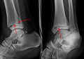
A triplane fracture of the ankle as seen on plain X-ray
-
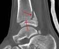
A triplane fracture of the ankle as seen on CT
-

A triplane fracture of the ankle as seen on CT
Top of page
Classification
There are several classification schemes for ankle fractures:
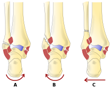
- The Lauge-Hansen classification categorises fractures based on the mechanism of the injury as it relates to the position of the foot and the deforming force (most common type is supination-external rotation)
- The Danis-Weber classification categorises ankle fractures by the level of the fracture of the distal fibula (type A = below the syndesmotic ligament, type B = at its level, type C = above the ligament), with use in assessing injury to the syndesmosis and theinterosseous membrane
- The Herscovici classification categorises medial malleolus fractures of the distal tibia based on level.
- The Ruedi-Allgower classification categorizes pilon fractures of the distal tibia.
Top of page
Treatment

Treatment of ankle fractures is dictated by the stability of the ankle joint. Certain fracture patterns are deemed stable, and may be treated similar to ankle sprains. All other types require surgery, most often an open reduction and internal fixation(ORIF), which is usually performed with permanently implanted metal hardware that holds the bones in place while the natural healing process occurs. A cast or splint will be required to immobilize the ankle following surgery.
|
Page Content |